Ultraviolet-visible Spectrophotometer application field
The simple principle of a Spectrophotometer
The Spectrophotometer uses a light source that can generate multiple wavelengths, through a series of spectroscopic devices, to generate a light source with a specific wavelength. After the light source passes through the test sample, part of the light source is absorbed, and the absorbance value of the sample is calculated, which is converted into the concentration of the sample. . The absorbance of a sample is directly proportional to the concentration of the sample.

Nucleic acid quantification:
Nucleic acid quantification is the most frequently used function of a Spectrophotometer . Can quantify buffer-soluble oligonucleotides, single- and double-stranded DNA, and RNA. The absorption wavelength of the highest absorption peak of nucleic acid is 260 nm. Each nucleic acid has a different molecular composition and therefore a different conversion factor. To quantify different types of nucleic acids, the corresponding coefficients must be selected in advance. For example: 1OD absorbance is equivalent to 50μg/ml dsDNA, 37μg/ml ssDNA, 40μg/ml RNA, 30μg/ml Olig. The absorbance value after the test is converted by the above coefficients to obtain the corresponding sample concentration. Before testing, select the correct program, enter the volume of the original solution and the dilution solution, and then test the blank solution and sample solution. However, the experiment was not all smooth sailing. Erratic readings are probably the biggest headache for experimenters. The more sensitive the instrument, the greater the absorbance drift will be.
In fact, the design principle and working principle of the Spectrophotometer allow the absorbance value to change within a certain range, that is, the instrument has a certain degree of accuracy and precision. Such as the accuracy of Eppendorf Biophotometer ≤ 1.0% (1A). It is normal for the results of such multiple tests to fluctuate between the average value of about 1.0%. In addition, it is also necessary to consider the physical and chemical properties of the nucleic acid itself and the pH value and ion concentration of the buffer in which the nucleic acid is dissolved: during the test, if the ion concentration is too high, it will also cause reading drift. Buffers, such as TE, can greatly stabilize the reading. The dilution concentration of the sample is also a factor that cannot be ignored: due to the inevitable existence of some fine particles in the sample, especially the nucleic acid sample. The presence of these small particles interferes with the test results. In order to minimize the impact of particles on the test results, it is required that the nucleic acid absorbance value is at least greater than 0.1A, and the absorbance value is preferably 0.1-1.5A. In this range, the interference of particles is relatively small and the result is stable.
This means that the concentration of the sample cannot be too low, or too high (beyond the test range of the photometer). Finally, there are operational factors, such as sufficient mixing, otherwise the absorbance value will be too low, or even negative; there should be no air bubbles in the mixed solution, and no suspended matter in the blank solution, otherwise the readings will drift drastically; the same cuvette should be used to test the blank solution and the sample , otherwise the concentration difference is too large; the conversion factor is consistent with the sample concentration unit; cuvettes with worn windows cannot be used; the volume of the sample needs to meet the minimum volume required by the cuvette and other operational matters.
In addition to the nucleic acid concentration, the Spectrophotometer also displays several very important ratios to indicate the purity of the sample, such as the ratio of A260/A280, which is used to evaluate the purity of the sample, because the absorption peak of the protein is 280nm. For pure samples, the ratio is greater than 1.8 (DNA) or 2.0 (RNA). If the ratio is lower than 1.8 or 2.0, it indicates the presence of protein or phenolic substances. A230 indicates that there are some pollutants in the sample, such as carbohydrates, polypeptides, phenol, etc., and the ratio of A260/A230 of relatively pure nucleic acids is greater than 2.0. The A320 detects the turbidity and other interfering factors of the solution. For pure samples, A320 is generally 0.
Direct quantification of protein (UV method):
This method is to directly test the protein at a wavelength of 280nm. Select the Warburg formula, the photometer can directly display the concentration of the sample, or choose the corresponding conversion method to convert the absorbance value into the concentration of the sample.
The protein determination process is very simple, first test the blank solution, and then directly test the protein. Due to the existence of some impurities in the buffer, it is generally necessary to eliminate the "background" information of 320nm, and set this function to "ON". Similar to testing nucleic acids, the absorbance value of A280 is required to be at least greater than 0.1A, and a good linear range is between 1.0-1.5. When choosing the Warburg formula to display the sample concentration in the experiment, it was found that the reading "drifted". This is a normal phenomenon. In fact, as long as the variation range of the absorbance value of A280 is not more than 1%, it shows that the result is very stable.
The reason for the drift is that the absorbance value of the Warburg formula is converted into concentration and multiplied by a certain coefficient. As long as there is a slight change in the absorbance value, the concentration will be amplified, which makes the result very unstable.
The direct protein quantification method is suitable for testing purer proteins with relatively single components. Compared with the colorimetric method, the ultraviolet direct quantitative method is fast and easy to operate; but it is easily interfered by parallel substances, such as DNA; in addition, the sensitivity is low and requires a higher concentration of protein.
Colorimetric protein quantification:
Protein is usually a compound of various proteins. The basis of colorimetric determination is protein composition: amino acids (such as tyrosine, serine) react with external chromogenic groups or dyes to produce colored substances. The concentration of colored substances is directly related to the number of amino acids reacted by the protein, thus reflecting the protein concentration.
Colorimetric methods generally include BCA, Bradford, Lowry and other methods.
Lowry method: Based on the earlier Biuret reaction and improved. The protein reacts with Cu2+ to produce a blue reactant. But compared with Biuret, Lowry's method is more sensitive. The disadvantage is that several different reagents need to be added sequentially; the reaction takes a long time; it is easily affected by non-protein substances; proteins containing EDTA, Tritonx-100, ammoniasulfate and other substances are not suitable for this method.
BCA (Bicinchoninineacidaassay) method: This is a newer and more sensitive protein test method. The protein to be analyzed reacts with Cu2+ in alkaline solution to produce Cu+, which forms a chelate with BCA to form a purple compound with an absorption peak at 562nm. This compound has a strong linear relationship with protein concentration, and the compound formed after the reaction is very stable. Compared with the Lowry method, the operation is simple and the sensitivity is high. However, similar to the Lowry method, it is susceptible to interference between proteins and detergents.
Bradford method: The principle of this method is that the protein reacts with Coomassie Brilliant Blue, and the resulting colored compound absorbs at 595nm. Its notable features are that the sensitivity is good, twice that of the Lowry and BCA test methods; the operation is simpler and the speed is faster; only one reaction reagent is needed; the compound can be stable for 1 hour, which is convenient for the result; and it is compatible with a series of Compatible with reducing agents (such as DTT, mercaptoethanol) that interfere with the Lowry, BCA reaction. However, it is still sensitive to detergents. The main disadvantage is that different standard products will lead to large differences in the results of the same sample, which is not comparable.
Some researchers who are new to the colorimetric method may be confused by the inconsistency of the results obtained by various colorimetric methods. Which method should they believe? Since the reacting groups and chromogenic groups of various methods are different, the sample concentration obtained by using several methods at the same time for the same sample is incomparable. For example: Keller et al. tested the protein in human milk. As a result, the concentration of Lowry and BCA was significantly higher than that of Bradford, and the difference was significant. Even if the same sample is measured, the standard samples selected by the same colorimetric method are inconsistent, and the concentrations after the test are also inconsistent. For example, if Lowry is used to test the protein in the cell homogenate, BSA is used as the standard product with a concentration of 1.34 mg/ml, and a globulin is used as the standard product with a concentration of 2.64 mg/ml. Therefore, before choosing a colorimetric method, it is best to refer to the chemical composition of the sample to be tested and find a standard protein with a similar chemical composition as a standard. In addition, the colorimetric method to quantify protein often has the problem that the absorbance value of the sample is too low, resulting in a large gap between the measured sample concentration and the actual concentration. The key issue is that the color after reaction has a certain half-life, so each colorimetric method lists the reaction test time, and all samples (including standard samples) need to be tested within this time. If the time is too long, the obtained absorbance value becomes smaller, and the converted concentration value decreases. In addition, the reaction temperature, the pH value of the solution, etc. are all important reasons that affect the experiment. In addition, it is very important to remember: it is better to use plastic colorimetry. Avoid using cuvettes made of quartz or glass, because the color after the reaction will stain the quartz or glass, resulting in inaccurate sample absorbance values.
Bacterial cell density (OD600):
The laboratory determines the growth density and growth period of bacteria, and infers the growth density of bacteria based on experience and visual inspection. When encountering more demanding experiments, it is necessary to use a Spectrophotometer to accurately measure the bacterial cell density. OD600 is the standard method for tracking microbial growth in liquid cultures. The culture solution without adding bacteria solution was used as the blank solution, and then the culture solution containing bacteria after culture was quantified. In order to ensure proper operation, a calibration curve needs to be generated for each microorganism and each instrument for cell counting with a microscope. Occasionally, the OD value of the bacterial solution will be negative in the experiment, because a color-developing medium is used, that is, after the bacteria are cultured for a period of time, they react with the medium and undergo a discoloration reaction. In addition, the north and south tides remind you: the test sample cannot be centrifuged to keep the bacteria in suspension.
An important accessory of the Spectrophotometer - cuvette:
Cuvettes are roughly divided into quartz cups, glass cups and plastic cups according to their materials. According to different measurement volumes, there are cuvettes and capillary cuvettes. Generally, quartz cups or glass cups are used to test nucleic acid and UV quantitative protein, but they are not suitable for colorimetric determination. Disposable plastic cups are required because the dyes in the reaction (such as Coomassie Brilliant Blue) can stain both quartz and glass. Plastic cups are generally not suitable for testing samples in the UV range.


- 1Application of Flame Atomic Absorption Spectrophotometry in Detection of Food Talc Powder
- 2Talking about the performance test of epoxy resin film coated on PET
- 3Spectrophotometer principle, structure and application do you know geometry?
- 4Principle, characteristics and application of ultraviolet Spectrophotometer
- 5Spectrophotometer Color Measurement Fundamentals
- 6Differences between visible, ultraviolet-visible, infrared, fluorescent and atomic absorption Spectrophotometer s
- 7Detection of nicotine content by Spectrophotometer
- 8How to detect the color difference of fluorescent color-changing materials?
- 9Liquid color difference detection solution
-
![YueFeng 752N Ultraviolet-visible Spectrophotometer]()
-
![YueFeng 721 Single beam of light Visible Spectrophotometer Test Transmittance/Absorbance]()
-
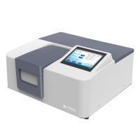
![JINGHUA H300 Dual beam of light Spectrophotometer Wavelength range 190nm-1100nm]()
-
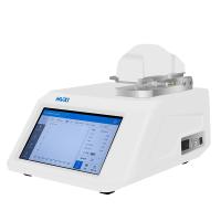
![HUXI Nano-900 UV-Visible, ultra-micro Spectrophotometer Light spectrum instrument]()
-
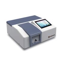
![JINGHUA JH752 Benchtop UV-Visible Spectrophotometer Bandwidth 4nm]()
-
![YUEFENG FP6428 Flame photometer Na/Ca]() YUEFENG FP6428 Flame photometer Na/Ca$ 1450.00
YUEFENG FP6428 Flame photometer Na/Ca$ 1450.00