What detection instrument is used for infrared spectroscopy testing?
Infrared spectrometers are usually Fourier transform infrared spectrometers (FTIR) which are sensitive and versatile instruments controlled by a computer. Fourier transform infrared spectrometers have high energy throughput and can generate useful spectra in seconds. Their wavelength or frequency calibration is accurate. All references to IR spectrometers in this chapter are references to FTIR spectrometers.
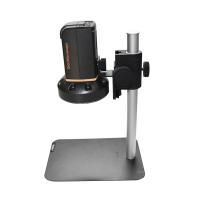
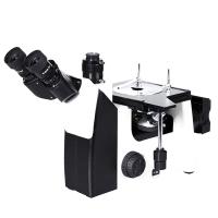
infrared microscope
Infrared microscopy is an excellent method for analyzing small samples or examining small areas of coatings. For example, this may help determine the cause of a failure or defect. Infrared microscopy combines an optical microscope adapted to infrared radiation and an infrared spectrometer. They can be an accessory to an infrared spectrometer or a stand-alone instrument. A means of visual observation of the sample is provided so that the analyst can correctly locate small samples or select desired portions from large samples. These means can be eyepieces and lenses for visual observation, or video cameras with suitable optics, and the images can appear on the monitor of a PC. With some microscopes, a computer can be used to control the microscope, selecting points, areas or lines to scan. Either transmission or reflection spectra are possible. For transmission measurements, a common problem is excessive sample thickness. The considerable pressure achieved by diamond cells can reduce many samples to the thickness required for useful spectra. Objectives using Attenuated Total Reflectance (ATR) technology are also available. Forensic applications, which may require analyzes like identifying small paint chips, rely heavily on infrared microscopy. Many analysts use infrared microscopy for most solid samples.
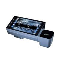
infrared imaging
Imaging is a growing trend in many areas of chemical analysis,12 as is in infrared spectroscopy. Just as cameras and microscopes produce visible images, it is possible to produce images with infrared radiation. For visible images, color is often used to distinguish different parts of the image. An infrared image has two-dimensional spatial information, as well as spectral information for each point on the image. By associating different colors with different parts of the spectrum, it is possible to create a visible image that contains information about the infrared response of the sample. For example, an image showing how a particular ester is distributed on a coating can be produced.
Early infrared imaging used a microscope with a stage that moved the sample past the infrared objective. As each point of the sample passes through the lens, an infrared spectrum is obtained. The resulting data will be a set of spectra, collected in a grid-like pattern over one region of one sample. A newer development is the infrared array Detector, which can have 256*256 or more Detectors arranged together. Each Detector in the array can produce a complete spectrum and thus can be scanned simultaneously to produce a complete image. This greatly reduces the time required to generate images and introduces the possibility of kinetic experiments, such as observing how a coated surface reacts or changes over time. Most imaging Detectors are integrated into infrared microscopes, although devices for macroscopic samples are available. Imaging Detectors may not be able to collect data over the broad wavelength range that other Detectors do, so the suitability of the Detector for the analyte of interest should be checked.


- 1Microscope classification, operation and application
- 2How to use trinocular Microscope
- 3How to detect the color difference of fluorescent color-changing materials?
- 4How is the content of non-fibroblasts determined?
- 5 How to measure cell wall thickness and cell cavity diameter?
- 6Determination of Fiber Length How is the distribution frequency calculated?
- 7How to determine the fiber morphology of plant fiber raw materials?
- 8Important instruments in clinical laboratories
- 9Coating Thickness Gauge: How to measure the dry film thickness of non-metallic substrates?