
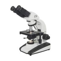
Varnishing XSP-BM-1CA Microscope Magnification 40X-1600X
SE XSP-BM-1CA

-
 Varnishing XSP-BM-20 Microscope Magnification 40X-1600X$ 1177.00SE
 -
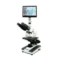 Varnishing XSP-BM-8CAP Tablet Microscope with 9.7 inch large screen tablet$ 1843.00
 -
 Varnishing XSP-BM-8C Microscope Magnification 40X-1600X$ 623.00SE
 -
 VARNISING XSP-BM-2CBAP Video Microscope with 9-inch large screen display$ 710.00SE


SHSGBM XSP-BM-1CA MicroscopeSPEC
SHSGBM XSP-BM-1CA MicroscopeDetails
SHSGBM XSP-BM-1CA MicroscopePacking list
- SKU
- NB044934
- Mechanical barrel length
- 160mm
- magnification
- 40X-1600X
- Stage size
- Mobile platform 125 × 110mm; moving range: 60 × 30mm, cursor: 0.1mm
- Focusing device
- 12Mm coarse fretting focusing coaxial mechanism, fine-tuning grid value 0.002mm
- condenser
- N.A.1.25 Adjustable medium Abbe condenser with variable light bar
- Color filter
- Blue, yellow, green
- Light source
- Incandescent lamp, 220V/20W, Light source adjustable

Introduction
The XSP-BM-1CAY biological microscope uses the magnification technology of an optical microscope to magnify and observe specimens made from small samples collected. It can be widely used in pathological examination, routine laboratory testing and teaching research.
This instrument uses a light source, the illumination can continuously adjust the brightness, the hinged lens barrel is tilted 45 degrees, and the eyepiece barrel can rotate 360 degrees freely.
Features
Real-time video observation: A video biomicroscope can capture real-time video images of biological samples through a camera, enabling users to continuously observe dynamic processes in the sample.
Digital imaging: Equipped with a digital imaging system, by connecting to a computer or other digital equipment, the image can be digitized for easy recording, storage and sharing.
High-resolution: High-resolution cameras ensure that details of tiny structures such as cells and microorganisms are clearly visible in the video.
Video recording and playback: With the video recording function, users can record observed videos for subsequent analysis, teaching or display, and support playback functions.
Autofocus and Contrast Adjustment: Some models are equipped with autofocus and contrast adjustment functions to improve observation and reduce the burden on the user.
Standard configuration
Eyepiece:
| category | Magnification | Field of view diameter |
| Plan eyepiece | 10X | φ18mm |
| 16X | φ11mm |
Objective:
| category | Magnification | Numerical aperture | Working distance |
| objective | 3w | 0.10NA | 37.5mm |
| 10X | 0.25NA | 7.31mm | |
| 40X | 0.65NA | 0.63mm | |
| 100X (oil) | 1.25NA | 0.18mm |
- 1Types of microscopes and common problems
- 2Overview of the different types of microscopes
- 3How does a digital microscope work?
- 4Microscope classification, operation and application
- 5How to use trinocular Microscope
- 6Microscope: a weapon to reveal the mysteries of microscopic life
- 7What are the defects of optical microscopes? Industry common sense
- 8Explanation of common terms for microscopes
- 9Characteristics of Electron Microscopes and Differences Between Several Microscopes
- 10Working Principle and Application Analysis of Confocal Microscope
- 1GB/T 44268.2-2024《Microscopes—Definition and measurement of illumination properties—Part 2:Illumination properties related to the colour in bright field microscopy》
- 2GB/T 44268.1-2024《Microscopes—Definition and measurement of illumination properties—Part 1: Image brightness and uniformity in bright field microscopy》
- 3GB/T 44276.2-2024《Microscopes—Cover glasses—Part 2:Quality of materials, standards of finish and mode of packaging》
- 4GB/T 44276.1-2024《Microscopes—Cover glasses—Part 1:Dimensional tolerances, thickness and optical properties》
- 5GB/T 44293-2024《Image measurement method for numerical aperture of microscopic objective》
- 6GB/T 39077-2024《Test methods for detecting detrimental phase in austenitic-ferritic (duplex) stainless steels》
- 7GB/T 43846.1-2024《Microscopes—Designation of microscope objectives—Part 1: Flatness of field/Plan》
- 8GB/T 43846.2-2024《Microscopes—Designation of microscope objectives—Part 2: Chromatic correction》
- 9GB/T 43846.3-2024《Microscopes—Designation of microscope objectives—Part 3: Spectral transmittance》
- 10GB/T 13777-2024《Test method for maturity of cotton fibers—Microscopic method》