
ShangGuang BM-13C Trinocular Epifluorescence Microscope
 BM-13C


ShangGuangLiuChang BM-13C Trinocular Epofluorescence MicroscopeSPEC
ShangGuangLiuChang BM-13C Trinocular Epofluorescence MicroscopeDetails
ShangGuangLiuChang BM-13C Trinocular Epofluorescence MicroscopePacking list
- SKU
- NB031640
- eyepiece
- WF10X / f20
- objective lens
- Flat-field color difference objective: 4 ×/0.10, 10 ×/0.25, 40 ×/0.65, 100 ×/1.25 (oil)
- Trinocular observation head
- Hinged 30 ° tilt
- pupil distance
- 55mm-75mm
- Mechanical barrel length
- 160mm
- Total magnification
- 40X~1000X
- converter
- Four hole converter
- stage
- Double-layer mechanical mobile (size: 160mmX140mm, moving range: 75mmX50mm)
- Focusing system
- Coarse micro-motion coaxial focusing, coarse elastic adjustable, with locking and limiting device plate, micro-motion grid value: 2μm
- Fluorescence filter set
- B (blue excitation) 420~485nm, G (green excitation) 460~550nm
- Mercury lamp box
- 100W/DC
- Fluorescence device power box
- AC: 110V or 220V
- light source
- 6V 20W halogen lamp, brightness adjustable
- condenser
- 1.25NA Abbe condenser, variable light bar
- Instrument Gross Weight
- 10kg
- Instrument size
- 21X50X54(cm)
- Packaging Dimensions
- 45X37X47(cm)

Features
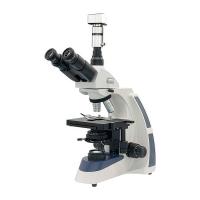
Trinocular fluorescence microscope XSP-BM13C is used for fluorescence microscopy and transmission field of view observation. It can clearly observe and identify chromosome specimens that are difficult to observe with ordinary microscopes. The perfect instrument for work. It can be used by departments such as scientific research, universities, medical care and epidemic prevention. Total magnification: 40X-1000X
Features
1. Equipped with wide-field eyepieces and fluorescent objective lenses, the field of view is flat and clear.
2. The planetary coaxial coarse and fine focus adjustment device is adopted, and the focus is comfortable and stable.
3. Two optical path outputs, one for observation and one for connecting camera.
4. A double-layer mechanical stage with graduated scales is adopted, and the handwheel is adjusted vertically and horizontally on the same axis, making the operation more convenient.
5. The transmission lighting adopts 6V 20W halogen lamp, and the brightness is adjustable; the fluorescent epi-illumination adopts 100W high-pressure mercury lamp, and an external power box is installed
optional parts
| Plan eyepiece | WF16X, WF20X |
| Micrometer eyepiece: 0.1 ruler shape, 0.05 ruler shape, 0.5 mesh shape, 0.2 mesh shape, 0.1 coordinate shape | |
| objective lens | Plan achromatic objective lens: 20X, 60X, 80X |
| Fluorescent Power Box | 100W wide Voltage constant power fluorescent power box with timer |
| epigraph fluorescence device | Fluorescence filter set: UV excitation light band 330~400nm, V excitation light band 395~415nm |
| Phase contrast device | Phase contrast objectives: 10X, 20X, 40X, 100X PHP |
| Plug-in phase-contrast condenser, pull-plate phase-contrast condenser, centering telescope | |
| ruler | Objective micrometer 0.01mm |
| Eyepiece micrometer: 0.1 ruler shape, 0.05 ruler shape, 0.5 mesh shape, 0.2 mesh shape, 0.1 coordinate shape | |
| camera port | Camera interface: 1X, 0.5X |
| Digital camera interface: 1X, 2.5X/4X | |
| Camera | ①Japan JVC camera + image acquisition card |
| With single, timing, continuous image acquisition, multimedia connection, printout and other functions | |
| ②Digital camera 1.3 million, 2 million, 3.2 million, 5 million pixels | |
| It has the functions of single, timing, and continuous image acquisition, display scale, measurement, etc., and can be connected to multimedia, printing, EMAIL and other ways of output. | |
| ③Digital camera + basic measurement management software | |
| 15 million pixel Canon SLR camera, with real-time image acquisition, image fusion, display scale, measurement and other functions, can be connected to multimedia, printing, EMAIL and other ways of output | |
| Image analysis software | ①Basic management software UV |
| Single sheet, timing, and continuous acquisition; scale and magnification can be displayed; length, angle, radius, perimeter, area, etc. can be measured for microscopic images; it can be connected to multimedia, print, EMAIL, etc. Output. | |
| ② Bioanalysis software UV-P | |
| Single sheet, timing, and continuous collection are available; rulers and magnifications can be displayed; various morphological analyzes of biological cells, germs, glands, etc. in the field of view can be performed, and output can be connected to multimedia, printing, and EMAIL. | |
| ③Particle size analysis software UV-G | |
| It can automatically measure and analyze the particles in the field of view, and output the distribution map and integral distribution map in a linear or nonlinear statistical manner according to the particle size, area, shape and other parameters of the particle, and can also obtain D10, D50, D90, etc. Common statistics. Can be connected to multimedia, printing, EMAIL and other ways of output. |
- 1Important instruments in clinical laboratories
- 2Fluorescence Microscope: a magical instrument that illuminates the microscopic world
- 3Microscope classification, operation and application
- 1GB/T 43846.1-2024《Microscopes—Designation of microscope objectives—Part 1: Flatness of field/Plan》
- 2GB/T 43846.2-2024《Microscopes—Designation of microscope objectives—Part 2: Chromatic correction》
- 3GB/T 43846.3-2024《Microscopes—Designation of microscope objectives—Part 3: Spectral transmittance》
- 4GB/T 13777-2024《Test method for maturity of cotton fibers—Microscopic method》
- 5GB/T 10561-2023《Determination of content of nonmetallic inclusions in steel—Micrographic method using standard diagrams》
- 6GB/T 42659-2023《Surface chemical analysis—Scanning probe microscopy—Determination of geometric quantities using SPM: Calibration of measuring systems》
- 7GB/T 42886-2023《Microscopes—Microscopes with digital imaging displays—Information provided to the user regarding imaging performance》
- 8GB/T 9247-2008《Microscopes - Condensers》
- 9GB/T 9246-2008《Microscopes - Oculars(eyepieces)》
- 10GB/T 22064-2008《Microscopes - Interfacing connection for 35 mm SLR photo cameras》